
Fig. 5
- ID
- ZDB-FIG-071127-15
- Publication
- Abe et al., 2007 - Function of FGF signaling in the developmental process of the median fin fold in zebrafish
- Other Figures
- All Figure Page
- Back to All Figure Page
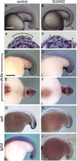
SU5402 did not affect dlx5a expression in the presumptive fin fold epidermis. Embryos were exposed to 0.17% DMSO for control (A, C, E, G, I) or 20 μM SU5402 (B, D, F, H, J) from 15 hpf for 3 h. Lateral view of embryos, anterior to the left. (a, b) HE staining of transverse sections at the level of each bar in panels A and B. Bracket in (a) indicates the area in which wedge-shaped cells were seen. The dorsal midline in the SU5402-treated embryo (b) was not covered with epidermis (arrowheads), and wedge-shaped cells for the fin fold were not formed. (C–F) In SU5402-treated embryos, dlx5a expression was retained as bilateral stripes in epidermal cells juxtaposed with the neural keel (D, F). (G–J) Expression patterns of sp9 (G, I) and fgf24 (H, J). Neither sp9 (H) nor fgf24 (J) was detected in the dorsal midline of SU5402-treated embryos. Bracket in (G, I) indicates sp9 expression observed in neural tissue. Scale bars in panels A and C are 200 μm. The panel width of (a, b) is 100 μm. |
| Genes: | |
|---|---|
| Fish: | |
| Condition: | |
| Anatomical Terms: | |
| Stage: | 14-19 somites |
Reprinted from Developmental Biology, 304(1), Abe, G., Ide, H., and Tamura, K., Function of FGF signaling in the developmental process of the median fin fold in zebrafish, 355-366, Copyright (2007) with permission from Elsevier. Full text @ Dev. Biol.