
Fig. 5
- ID
- ZDB-FIG-071025-5
- Publication
- Mann et al., 2007 - Vestigial-like-2b (VITO-1b) and Tead-3a (Tef-5a) expression in zebrafish skeletal muscle, brain and notochord
- Other Figures
- All Figure Page
- Back to All Figure Page
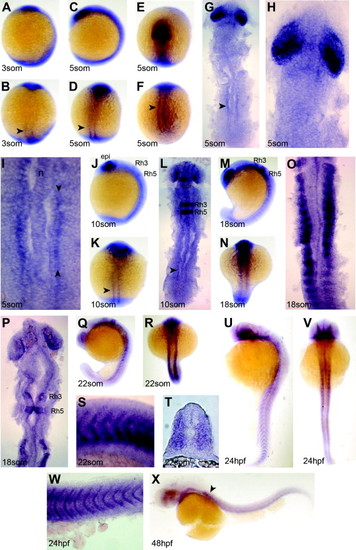
Developmental expression of tead-3a. In-situ mRNA hybridisation for tead-3a in lateral (A, C, J, M, Q, U, and X) and dorsal (B, D, K, N, R, and V) views of wholemount (A–F, J, K, M, N, Q, R, U, V, and X), flatmount (G–I, L, O, P, S, and W) or cryosection (T, dorsal to top). (A–L) Somitic expression begins at 3 som in narrow adaxial bands (arrowheads) on either side of the notochord (n) that extend into the anterior presomitic mesoderm (I). Strong retinal expression is evident from 5 som, with epiphysis (epi) and rhombomeres (Rh) expression apparent by 10 som. (M–T) Adaxial expression persists until differentiation of fast muscle begins at 18 som, when signal is detected more broadly throughout the somite (T), but strongest in a posterior–ventral domain (S). Epiphysis and rhombomere expression declines. (U–X) By the end of somitogenesis, tead-3a mRNA is concentrated at the somite borders and in the branchial region. |
| Gene: | |
|---|---|
| Fish: | |
| Anatomical Terms: | |
| Stage Range: | 1-4 somites to Long-pec |
Reprinted from Gene expression patterns : GEP, 7(8), Mann, C.J., Osborn, D.P., and Hughes, S.M., Vestigial-like-2b (VITO-1b) and Tead-3a (Tef-5a) expression in zebrafish skeletal muscle, brain and notochord, 827-836, Copyright (2007) with permission from Elsevier. Full text @ Gene Expr. Patterns