
Fig. 8
- ID
- ZDB-FIG-071004-30
- Publication
- Tyurina et al., 2005 - Zebrafish Gli3 functions as both an activator and a repressor in Hedgehog signaling
- Other Figures
- All Figure Page
- Back to All Figure Page
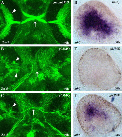
Gli3 is required for RGC differentiation. (A) At 40 h of development, large numbers of Zn-5 labeled retinal ganglion cells (RGCs) (arrowheads) in the eyes have differentiated and extended axons across the midline to form the optic nerve and chiasm (arrow). (B) In severely affected gli3MO injected embryos, very few RGCs differentiated, and those that were present were distributed aberrantly in the eye (arrowheads). (C) In less severely affected gli3MO injected embryos, reduced numbers of RGCs differentiated in the correct location and extended axons across the midline to form a thin optic nerve and chiasm (arrow). (D) At 34 h, the zebrafish atonal homolog ath5 is expressed throughout the central region of the differentiating retina. (E) In severely affected gli3MO-injected embryos, ath5 expression was completely eliminated in the eye. (F) In less severely affected gli3MO-injected embryos, ath5 expression was strongly reduced. (A–C) Fluorescent labeling of RGCs at 40 h using the Zn-5 antibody, ventral views of head, anterior up. (D–F) ath5 in situ labeling of the eyes at 34 h, lateral views of the left eye, dorsal up, anterior to the left. |
Reprinted from Developmental Biology, 277(2), Tyurina, O.V., Guner, B., Popova, E., Feng, J., Schier, A.F., Kohtz, J.D., and Karlstrom, R.O., Zebrafish Gli3 functions as both an activator and a repressor in Hedgehog signaling, 537-556, Copyright (2005) with permission from Elsevier. Full text @ Dev. Biol.