
Fig. 8
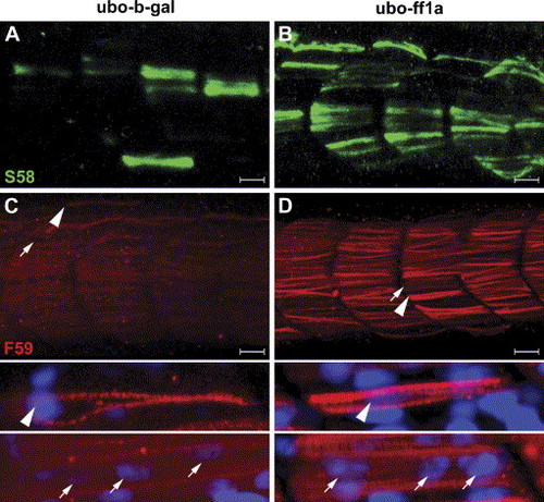
Partial restoration of muscle myofibril in ubo mutants by ectopic expression of ff1a. The ubo mutants were injected with (A, C) control β-gal or (B, D) ff1a RNA followed by staining with (A, B) S58 or (C, D) F59 antibodies at 24 hpf, and somites 14–17 are shown in lateral view with anterior towards the left. (A, B) Slow muscle specific staining of S58 confirmed that the differentiation of slow myofiber was partially restored by transient expression of ff1a in ubo mutants. (C) In the ubo mutants, very few myofibrils can be stained by F59. (D) Ectopic expression of ff1a resulted in the formation of more myofibrils at the surface. Arrowheads point to mono-nucleated myofibrils as shown in the enlarged figure below, including nuclear staining by DAPI. The arrows point to fast multi-nucleated myofibrils in the enlarged picture. The F59 antibody detects slow myofibrils strongly and fast myofibrils weakly. Scale bars are 20 μm. |
Reprinted from Developmental Biology, 286(2), Sheela, S.G., Lee, W.C., Lin, W.W., and Chung, B.C., Zebrafish ftz-f1a (nuclear receptor 5a2) functions in skeletal muscle organization, 377-390, Copyright (2005) with permission from Elsevier. Full text @ Dev. Biol.