
FIGURE
Fig. 3
- ID
- ZDB-FIG-070918-37
- Publication
- Pugacheva et al., 2006 - Cloning and characterization of zebrafish CTCF: Developmental expression patterns, regulation of the promoter region, and evolutionary aspects of gene organization
- Other Figures
- All Figure Page
- Back to All Figure Page
Fig. 3
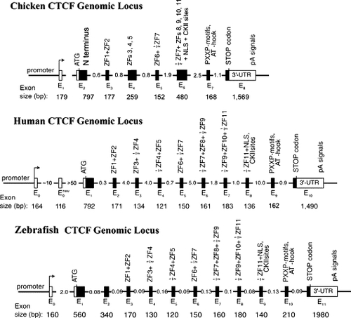
Genomic organization of the chicken, human and zebrafish CTCF genes. Filled boxes, protein coding exons; open boxes, untranslated exons; arrows, transcription start sites. The numbers indicate the estimated sizes of exons and introns in kilobases. The eleven zinc fingers of mammalian CTCF are distributed in exons E2 to E8, with several ZFs being split across neighboring exons. Structure of the avian and human CTCF genes is shown according to Ohlsson et al. (2001) |
Expression Data
Expression Detail
Antibody Labeling
Phenotype Data
Phenotype Detail
Acknowledgments
This image is the copyrighted work of the attributed author or publisher, and
ZFIN has permission only to display this image to its users.
Additional permissions should be obtained from the applicable author or publisher of the image.
Reprinted from Gene, 375, Pugacheva, E.M., Kwon, Y.W., Hukriede, N.A., Pack, S., Flanagan, P.T., Ahn, J.C., Park, J.A., Choi, K.S., Kim, K.W., Loukinov, D., Dawid, I.B., and Lobanenkov, V.V., Cloning and characterization of zebrafish CTCF: Developmental expression patterns, regulation of the promoter region, and evolutionary aspects of gene organization, 26-36, Copyright (2006) with permission from Elsevier. Full text @ Gene