
Fig. 6
- ID
- ZDB-FIG-070914-67
- Publication
- Steffen et al., 2007 - The zebrafish runzel muscular dystrophy is linked to the titin gene
- Other Figures
- All Figure Page
- Back to All Figure Page
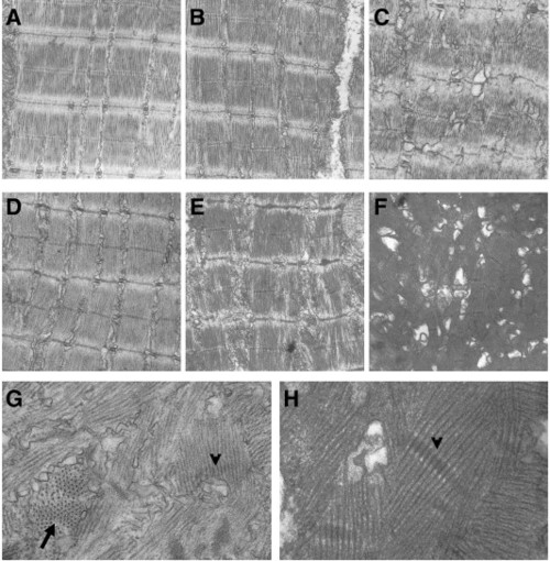
ruz mutants produce normal sarcomeres but show progressive sarcomeric misalignment. (A–H) Longitudinal sections of skeletal muscle from 3.5 dpf (A–C) or 6.5 dpf (D–H) fish were analyzed by transmission electron microscopy. (A) Wild-type fish at 3.5 dpf show normal sarcomeric organization and alignment. (B, C) ruz mutants contain some normal sarcomeres at 3.5 dpf (B) but are also beginning to show sarcomeric disruption (C). At 6.5 dpf, wild-type fish still display normal sarcomeres (D) while ruz mutants contain only rare regions of ordered myofibrils (E). Instead, myofibers contain collapsed sarcomeres with no apparent organization or alignment (F–H). Arrow indicates fibrils perpendicular to the plane of the section. Arrowheads indicate selected Z-disc structures. |
| Fish: | |
|---|---|
| Observed In: | |
| Stage Range: | Protruding-mouth to Day 6 |
Reprinted from Developmental Biology, 309(2), Steffen, L.S., Guyon, J.R., Vogel, E.D., Howell, M.H., Zhou, Y., Weber, G.J., Zon, L.I., and Kunkel, L.M., The zebrafish runzel muscular dystrophy is linked to the titin gene, 180-192, Copyright (2007) with permission from Elsevier. Full text @ Dev. Biol.