
FIGURE
Fig. 1
- ID
- ZDB-FIG-070914-63
- Publication
- Steffen et al., 2007 - The zebrafish runzel muscular dystrophy is linked to the titin gene
- Other Figures
- All Figure Page
- Back to All Figure Page
Fig. 1
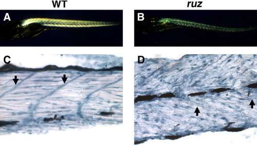
Skeletal muscle is severely disorganized in ruz mutants. (A) Wild-type fish skeletal muscle is highly birefringent at 5–6 dpf. (B) 5–6 dpf ruz homozygous mutants show decreased skeletal muscle birefringence and, on a TL background, body curvature. (C, D) Longitudinal sections of zebrafish skeletal muscle at 5 dpf were stained with hematoxylin. (C) Wild-type muscle shows myofiber alignment between myosepta and peripherally localized nuclei. (D) ruz muscle myofibers appear unaligned with abnormally shaped nuclei. Arrows indicate myosepta. |
Expression Data
Expression Detail
Antibody Labeling
Phenotype Data
| Fish: | |
|---|---|
| Observed In: | |
| Stage Range: | Day 5 to Day 6 |
Phenotype Detail
Acknowledgments
This image is the copyrighted work of the attributed author or publisher, and
ZFIN has permission only to display this image to its users.
Additional permissions should be obtained from the applicable author or publisher of the image.
Reprinted from Developmental Biology, 309(2), Steffen, L.S., Guyon, J.R., Vogel, E.D., Howell, M.H., Zhou, Y., Weber, G.J., Zon, L.I., and Kunkel, L.M., The zebrafish runzel muscular dystrophy is linked to the titin gene, 180-192, Copyright (2007) with permission from Elsevier. Full text @ Dev. Biol.