
Fig. S1
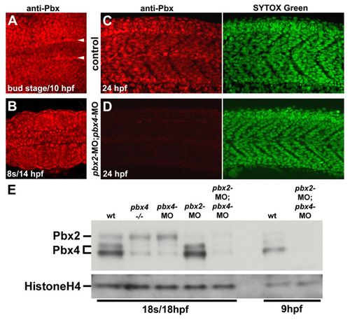
Pbx expression during early myogenesis stages is lost in pbx2-MO; pbx4-MO embryos. (A-D) Anti-zebrafish pan-Pbx staining in (A-C) whole wild-type control and (D) pbx2-MO; pbx4-MO embryos at (A) bud stage/10 hpf, (B) 10 somite stage (s)/14 hpf, or (C) 24 hpf. 24 hpf embryos were also stained with Sytox Green to identify nuclei. (A,B) Show dorsal views, anterior towards the left, of (A) early paraxial mesoderm and (B) somites 3-5. (A) Arrowheads label the rows of adaxial cells next to the notochord. (C,D) Show lateral views, anterior towards the left, centered on somites 8-11. (E) Western blot using zebrafish pan-Pbx antibody and Histone H4 antibody. Western analysis with a Pbx4-specific antibody identified the two Pbx4 bands (data not shown). |