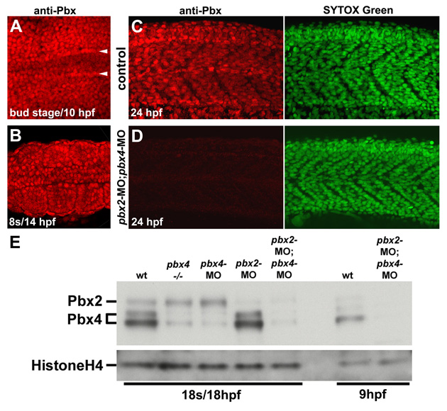
Fig. S1 Pbx expression during early myogenesis stages is lost in pbx2-MO; pbx4-MO embryos. (A-D) Anti-zebrafish pan-Pbx staining in (A-C) whole wild-type control and (D) pbx2-MO; pbx4-MO embryos at (A) bud stage/10 hpf, (B) 10 somite stage (s)/14 hpf, or (C) 24 hpf. 24 hpf embryos were also stained with Sytox Green to identify nuclei. (A,B) Show dorsal views, anterior towards the left, of (A) early paraxial mesoderm and (B) somites 3-5. (A) Arrowheads label the rows of adaxial cells next to the notochord. (C,D) Show lateral views, anterior towards the left, centered on somites 8-11. (E) Western blot using zebrafish pan-Pbx antibody and Histone H4 antibody. Western analysis with a Pbx4-specific antibody identified the two Pbx4 bands (data not shown).
Image
Figure Caption
Acknowledgments
This image is the copyrighted work of the attributed author or publisher, and
ZFIN has permission only to display this image to its users.
Additional permissions should be obtained from the applicable author or publisher of the image.
Full text @ Development

