
Fig. 3
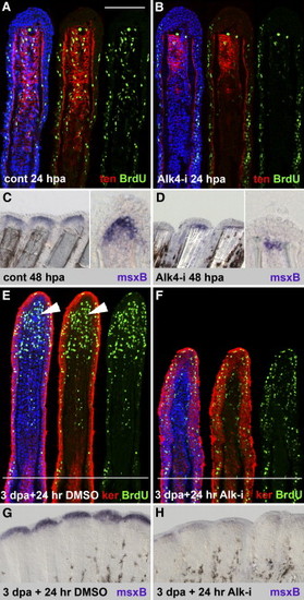
ActβA Signaling Is Required for Blastema Outgrowth and Maintenance (A and B) Longitudinal sections of the fin rays at 24 hpa, triply stained with BrdU antibody in green, anti-tenascin in red, and ToPro3 in blue. In the control, the enhanced number of BrdU-positive cells in the epidermis and mesenchyme indicates a proliferative phase of regeneration (A). The mesenchymal domain with proliferating cells overlaps with the tenascin domain. Fins treated with 10 μM Alk4-i contain an enhanced number of BrdU-positive cells in the epidermis, but mesenchymal proliferation is decreased (B). The extent of tenascin domain is reduced to half size when compared to control (two to three representative sections of four fins were analyzed). The scale bar in (A) represents 100 μm. (C and D) In situ hybridization of fins with msxB antisense probe. At 48 hpa, control fins display strong msxB expression distally from the amputation plane in the blastema, as seen on a whole mount (left panel) and a longitudinal section (right panel) (C). Inhibition of ActβA completely depletes msxB (35% of the rays in five fins) or reduces msxB expression to a single row of mesenchymal cells that remain at the amputation plane (65% of the rays in five fins) (D). (E and F) Longitudinal sections of the fin rays at 4 dpa (3 days at normal conditions and 24 hr with 0.1% DMSO) triply stained with BrdU antibody in green, anti-pan-keratin in red, and DAPI in blue. The white lines demarcate the amputation plane. In control fins, the blastema (arrowheads) displays a massive proliferation, as visualized by BrdU incorporation, and no keratin staining (E). The intensity of keratin is correlated with mesenchymal differentiation: high levels in the proximal mesenchyme (above the amputation plane) and no staining in the blastema. Keratin staining strongly labels the epidermis. Fins regenerating in normal conditions for 3 days and then transferred to water with Alk4-i for the next 24 hr (drug-shift experiment) are shown (F). The smaller size of the outgrowth and reduction of blastemal proliferation demonstrates a block of normally initiated regeneration after the exposure to the drug. Two to three representative sections of four fins were analyzed. (G and H) In situ hybridization of fins with msxB antisense probe. In control fins at 4 dpa (3 days at normal conditions and 24 hr with 0.1% DMSO), msxB mRNA is detected in the blastema of the outgrowth (G). msxB expression is strongly diminished in fins exposed to Alk4-i for 24 hr starting at 3 dpa (four out of six fins). (H) |