
Fig. 5
- ID
- ZDB-FIG-070815-17
- Publication
- Schibler et al., 2007 - A screen for genetic defects of the zebrafish ear
- Other Figures
- All Figure Page
- Back to All Figure Page
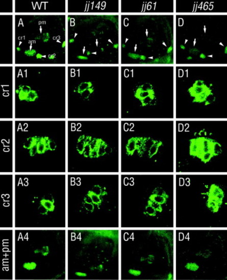
Hair cell morphology and distribution in otolith mutants. Hair cells were evaluated by HCS-1 antibody staining at 5 dpf. Images of whole ears in wild-type and mutant animals are shown in the top row. Panels below, in the rows labeled cr1, cr2, and cr3, show enlargements of anterior, lateral, and posterior cristae, respectively. The bottom row, labeled am + pm, shows the anterior and the posterior macula. No otolith mutants show obvious defects of hair cell distribution. In (A–D), cristae are indicated with arrowheads, and maculae with arrows. The following structures are labeled in (A): cr1, anterior crista; cr2, lateral crista; cr3, posterior crista; am, anterior macula; and pm, posterior macula. In all panels, dorsal is up and anterior is left. |
| Fish: | |
|---|---|
| Observed In: | |
| Stage: | Day 5 |
Reprinted from Mechanisms of Development, 124(7-8), Schibler, A., and Malicki, J., A screen for genetic defects of the zebrafish ear, 592-604, Copyright (2007) with permission from Elsevier. Full text @ Mech. Dev.