
Fig. 4
- ID
- ZDB-FIG-070815-16
- Publication
- Schibler et al., 2007 - A screen for genetic defects of the zebrafish ear
- Other Figures
- All Figure Page
- Back to All Figure Page
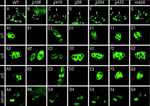
Hair cell distribution in mutants of ear morphology. Hair cells were evaluated by HCS-1 antibody staining at 5 dpf. Images of whole ears in wild-type and mutant animals are shown in the top row. Panels below, rows labeled cr1, cr2, and cr3, represent enlargements of anterior, lateral, and posterior cristae, respectively. The bottom row, labeled am + pm, shows the anterior and the posterior macula. The most severe phenotypes are observed in mikre uchojj108, ale uchojj410, and hako mimijj59 mutant animals. In mikre uchojj108, only a rudiment of one crista appears to be present (B2) and both maculae are severely misshapen (B4). A subpopulation of ale uchojj410 mutants exhibit disorganized and expanded cristae (C3). hako mimijj59 mutants lack both anterior and posterior cristae but retain the wild-type appearance of maculae (D4) and the lateral crista (D2). The remaining mutants do not display obvious defects of sensory patch morphology or positioning. In panels (A–G), cristae are indicated with arrowheads and maculae with arrows. The following structures are labeled in (A): cr1, anterior crista; cr2, lateral crista; cr3, posterior crista; am, anterior macula; and pm, posterior macula. In all panels, dorsal is up and anterior is left. |
| Fish: | |
|---|---|
| Observed In: | |
| Stage: | Day 5 |
Reprinted from Mechanisms of Development, 124(7-8), Schibler, A., and Malicki, J., A screen for genetic defects of the zebrafish ear, 592-604, Copyright (2007) with permission from Elsevier. Full text @ Mech. Dev.