|
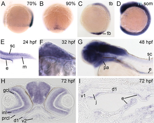
rerea is expressed dynamically in all developmental stages examined. All panels show whole-mount in situ hybridizations of rerea for wild-type embryos or larvae of ages indicated in upper right corner. A-G: Lateral views with anterior to the left (A,C,D,G), dorsal view with animal pole up (B), lateral view on tail (E), and frontal view of fin bud with medial to the left (F). H,I: Frontal section though head (H) and horizontal sections with anterior to the left through the pharyngeal arches (I). d1, dorsal cartilages of arch 1; e, endoderm; gcl, ganglion cell layer; inl, inner nuclear layer; j, joint; m, somitic mesoderm; pa, pharyngeal arches; prcl, photoreceptor cell layer; sc, spinal cord; tb, tail bud; v1 and v2, ventral cartilages of arches 1 and 2, respectively.
|