
Fig. 4
- ID
- ZDB-FIG-070713-4
- Publication
- Kida et al., 2007 - Daam1 regulates the endocytosis of EphB during the convergent extension of the zebrafish notochord
- Other Figures
- All Figure Page
- Back to All Figure Page
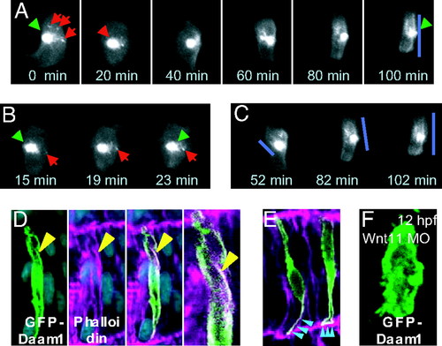
Subcellular localization of Daam1 in notochord cells. (A) Serial pictures of a notochord cell taken at 20-min intervals. EGFP-Daam1-positive vesicles (red arrows) moved from the caudal cell surface to the center (green arrowhead). Daam1-positive fibers were formed on the caudal side of the cell (blue line). (B) Serial pictures of another notochord cell taken at 4-min intervals. EGFP-Daam1-positive vesicles (red arrows) moved from the caudal cell surface to the center (green arrowhead). (C) Serial pictures of the cell shown in (A). GFP-tagged Daam1 accumulated in the fibers near the caudal cell surface (blue bars). (D and E) Subcellular localization of GFP-tagged Daam1 is shown in green. At this stage, Daam1 colocalized with F-actin in the cell cortex and the fibers formed near the caudal surface (yellow arrowheads). Note that the colocalization is more evident at the lateral ends that make tight junctions with surrounding tissues (blue arrowheads in E). (F) When wnt11 MO was injected, the notochord cells did not elongate but, rather, formed small spiky protrusions where Daam1 colocalized. Note that Daam1 did not localize in the endocytic vesicles or the fibers in the cell cortex and, instead, stayed at the cell membrane. |
| Fish: | |
|---|---|
| Knockdown Reagent: | |
| Observed In: | |
| Stage: | 5-9 somites |