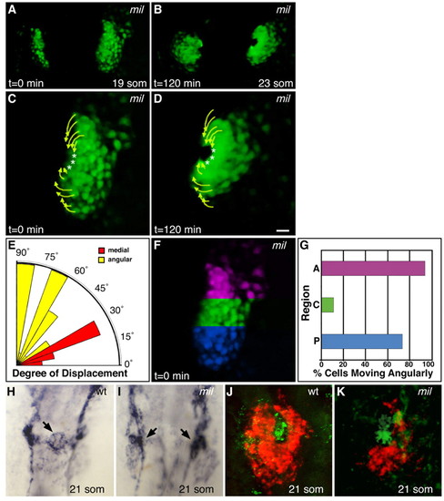
Angular cell movement occurs in the absence of initial medial movement. (A-D) Selected images from a time-lapse of cardiac fusion in a typical mil mutant zebrafish embryo expressing Tg(cmlc2:egfp) (see Movie 3 in the supplementary material), exhibiting cardiac morphology at the (A) 19-somite and (B) 23-somite stages. Dorsal views, anterior to the top. (C,D) Paths traveled by cardiomyocytes in the right lateral heart field during the entire time-lapse (arrows and asterisks as described for Fig. 1). Tracks displayed represent a subset of the tracked cells in this time-lapse. Although initial medial movement is lost, mil mutant cardiomyocytes exhibit a phase of angular movement. Images in C,D are double the magnification of those in A,B. Scale bar: 20 μm. (E) Radial bar graph (see Fig. 1J,L) depicting degree of displacement for all tracked mil mutant cardiomyocytes. Most cells exhibit angular movement. (F,G) Location of cardiomyocytes moving angularly in mil mutants. In mil mutants, as in wild-type embryos, angular movement is regionally restricted. See Table 1 for additional data. (H,I) In situ hybridization for flk1 (kdr - ZFIN) expression in wild-type (H) and mil mutant (I) embryos at the 21-somite stage. Dorsal views, anterior to the top; both images shown at the same magnification. Arrows indicate clusters of presumed endocardial precursors. (J,K) Two-color fluorescent in situ hybridization for expression of cmlc2 (red) and fli1a (green) in wild-type (J) and mil mutant (K) embryos at the 21-somite stage. Dorsal views, anterior to the top; only the right lateral heart field is shown in K; both images shown at the same magnification. In both wild-type and mil mutant embryos, the presumed endocardial precursors are clustered adjacent to the central cardiomyocytes.
|
