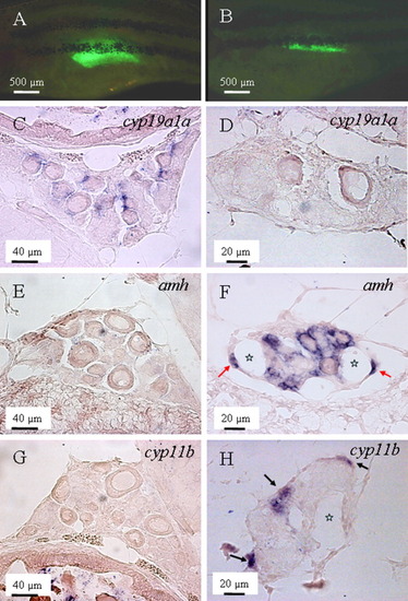
Expression pattern of cyp19a1a, amh, and cyp11b in the "normal" ovaries and transforming ovaries. A,C,E,G: The gonad of a female individual at 4 weeks postfertilization (wpf) of age is shown. B,D,F,H: The transforming gonad of a male individuals at 5 wpf of age is shown. A: An individual with "normal" ovary indicated by continuous accumulation of EGFP during the previous week. B: An individual with transforming ovary indicated by continuous decrease of enhanced green fluorescent protein (EGFP) during the previous week. C,D:cyp19a1a was expressed in the presumptive granulosa cells surrounding the normal oocytes (C), but not in the transforming ovary (D). E,F:amh initially could not be detected in the normal ovary (E), but became highly expressed in the cells surrounding the degenerating oocytes (F), and remained so even after the complete degeneration of oocytes (F, red arrow). G,H: Similarly to amh, cyp11b was also expressed during gonadal transformation. H: However, it was usually first expressed on the edge of the gonad (black arrows), without obvious correlation with the position of degenerating oocytes. Stars indicate the cavities after degeneration of oocytes.
|
