
Fig. 1
- ID
- ZDB-FIG-070613-2
- Publication
- Shin et al., 2007 - Notch signaling regulates neural precursor allocation and binary neuronal fate decisions in zebrafish
- Other Figures
- All Figure Page
- Back to All Figure Page
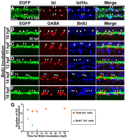
Neural plate olig2:EGFP+ precursors generate PMNs and KA' interneurons at the same time. (A) Dorsal view of the posterior neural plate at 11.5 hpf (five-somite stage) and (B-F) lateral view of 6- to 12-somite regions of spinal cord at 25 hpf in Tg(olig2:egfp) zebrafish embryos. (A) Heterogeneous primary neuronal populations within the EGFP+ domain. Asterisks and arrowheads mark EGFP+ Hu+ Isl+ PMNs and EGFP+ Hu+ Isl- interneurons, respectively. Isl protein is nuclear, whereas Hu is cytoplasmic. (B-F) Embryos were incubated with BrdU at successive timepoints (as shown to the left of each panel) and labeled with anti-GABA (red) and anti-BrdU (blue) antibodies. Arrows and arrowheads mark BrdU+ and BrdU- KA' interneurons, respectively. (B) Four KA' interneurons were formed from EGFP+ precursors that underwent S phase at 10 hfp (arrows). Arrowhead marks BrdU- KA' interneuron, indicating that the postmitotic cell was formed before or after 10 hpf. (C) Two BrdU+ KA' interneurons (arrows) and two BrdU- KA' interneurons (arrowheads) were detected in the embryos that were incubated with BrdU at 11 hpf. (D,E,F) Embryos incubated with BrdU at 12, 14 and 18 hpf. No BrdU+ KA' interneurons were evident. (G) Average of all KA' cells (squares) versus BrdU+ KA' cells (triangles) (n=4 animals each). S phase for the last KA' interneurons produced occurs between 10 and 12 hpf. Error bars represent s.e.m. Scale bar: 25 µm. |