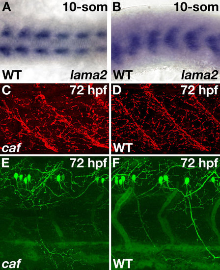
Expression of lama2 mRNA in the adaxial cells of a wild-type embryo and neuromuscular junction formation and primary motor neuron outgrowth. (A) In situ hybridization to Lama2 mRNA shows initial expression in the adaxial cells at the 10-somite stage. The adaxial cells are the first muscle cells of the embryo to differentiate. Wild-type, dorsal view, anterior to the left. (B) Wild type, lateral view, anterior to the left. (C) Staining with rhodamine-conjugated bungarotoxin allows visualization of the neuromuscular junctions (NMJs). No differences in the extent of NMJ formation were seen between caf embryos and wild-type siblings. Panels show confocal projections, lateral view, anterior to the left. Full projections are available as SI Movies 2 and 3. (D) Crossing caf zebrafish with the Tg(cmet:EGFP)ed6 line allowed visualization of the primary motor neurons. No defects in primary motor neuron formation or outgrowth were seen in caf embryos compared to wild-type siblings. Panels show confocal projections, lateral view, anterior to the left. Full projections are available as SI Movies 4 and 5.
|
