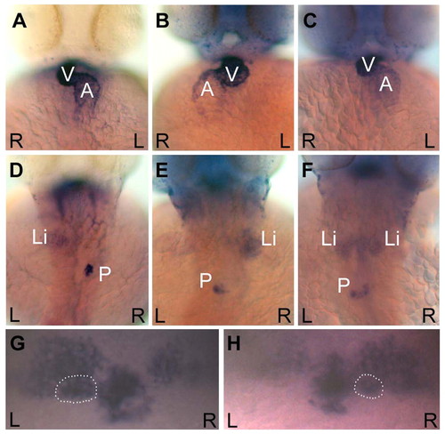
curly up embryos display defects in visceral and brain asymmetries. Ventral views of the heart (A-C) and dorsal views of the liver and pancreas (D-F) in cup embryos at 48 hpf. The heart, liver and pancreas were visualized by in situ hybridizations for cmlc2, fkd2, and ins, respectively. Wild-type organ positioning can be seen in approximately one third of a cup mutant population (A and D), where the ventricle loops to the right (A), and the liver resides on the left and the pancreas on the right (D). cup embryos also exhibit situs inversus, as seen by the complete reversal of the heart (B), liver and pancreas (E). Approximately one-third of cup mutants display heterotaxia. Included in this category was an embryo that had correct heart looping (C) but showed a duplicated liver with a reversed pancreas (F). (G,H) Dorsal views of the habenular nuclei and the pineal complex in 3-day cup embryos. The parapineal is outlined in each panel. (G) cup mutant with the wild-type pattern of more intense lov expression in the left habenula, and left parapineal placement visualized by otx5. (H) cup mutant with reversed diencephalic asymmetries. L, left; R, right; V, ventricle; A, atrium; Li, liver; P, pancreas.
|
