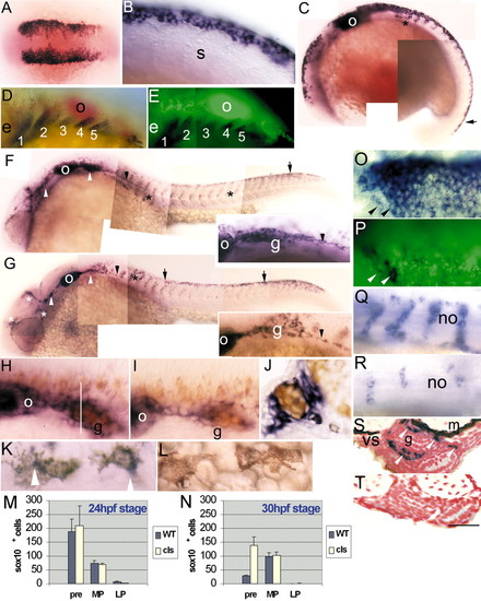
Embryonic sox10 expression in wild-type and cls embryos. (A,B) sox10 is expressed in most cranial (A; five-somite stage) and trunk (B; 14-somite stage) premigratory NCCs; double mRNA in situ hybridisation with fkd6 (O, purple) and sox10 (P, green) in a six-somite stage embryo reveals extensive overlap, although some individual NCCs lack sox10 (arrowheads). (C) 18-somite stage embryo shows strong expression in premigratory (black arrow) and migrating (asterisk) NCCs and otic vesicle (o). (D,E) Double mRNA in situ hybridisation with dlx2 (D, purple) and sox10 (E, green) in 29 hpf stage embryos shows absence of sox10 expression in developing branchial arches (1-5). (F) By 24 hpf in wild types, strong expression is associated with cranial ganglia (white arrowheads) and posterior lateral line nerve (black arrowhead; enlarged in inset), otic vesicle (o), migrating NCCs throughout trunk (asterisks) and in premigratory crest (arrow). (G) At 24 hpf in cls mutants, expression in the head is clustered (white asterisks) and cranial ganglia have reduced labelling (white arrowheads). Cells expressing sox10 extend along the posterior lateral line nerve (arrowhead and inset). Rostral trunk shows some migrating cells (black asterisk), but sox10-positive cells are clustered dorsal to the neural tube (arrows) in trunk and tail. (H,I) Combined sox10 in situ hybridisation (purple) and anti-Hu antibody labelling (orange) shows strong sox10 expression associated with wild-type (H) posterior lateral line ganglion (g), much reduced in cls mutant (I). (J) In transverse section of wild-type ganglion (approximate position indicated by white line in H), sox10 expression is strong peripherally (non-neuronal cells), but absent centrally (neurones). (K,L) 36 hpf wild-type embryos show weak expression in some melanophores, but not all. Thus, weak expression (arrowhead) is seen in some cells of the dorsal stripe (K), but not in cells on the yolk sac (L). (M,N) Number of sox10-expressing NCCs in different locations (premigratory, pre; migrating on medial pathway, MP; migrating on lateral pathway, LP) of trunk and anterior tail (somites 1-20) at 24 (M) and 30 hpf (N) in WT and cls mutants. (Q,R) Segmentally arranged lines of sox10-positive cells lying adjacent to the notochord (no), presumably glia, are abundant in wild type (Q) and only weakly affected in cls mutants (R) at 40 hpf. (S,T) Transverse section of trunk of 60 hpf wild-type embryo (S) shows enteric nervous system expression (arrowheads) lateral to the gut (g), absent in cls mutant (T). e, eye; m, muscle; s, somite; vs, ventral stripe melanophores. All images are lateral views, rostral towards the left, dorsal upwards, except dorsal views of A,K,O,P. Scale bar: 200 μm in A; 50 μm in B,H-I,K-L; 120 μm in C; 75 μm in D,E; 150 μm in F,G; 35 μm in J; 65 μm in O,P; 55 μm in Q,R; 45 μm in S,T.
|
