
Fig. 1
- ID
- ZDB-FIG-070314-40
- Publication
- Liu et al., 2007 - Notch signaling controls the differentiation of transporting epithelia and multiciliated cells in the zebrafish pronephros
- Other Figures
- All Figure Page
- Back to All Figure Page
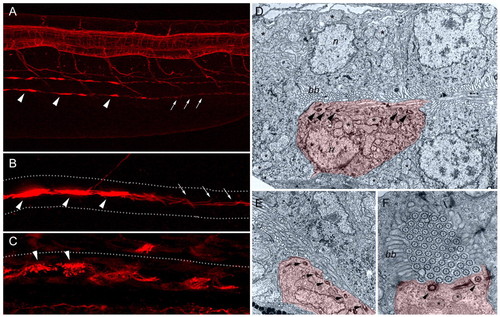
Distribution of multiciliated cells and transporting epithelia containing single cilia in the pronephros. (A) Whole-mount immunofluorescence of the trunk region of a 48 hpf embryo stained with anti-acetylated tubulin reveals bright cilia bundles (arrowheads) in the pronephric lumens, as well as single ciliated cells (arrows) in a more caudal nephron segment. (B) At higher magnification, compressed bundles of cilia (arrowheads) and single cilia (arrows) can be observed in the pronephric lumen. (C) Bundles of cilia emanating from individual cells project into a distended lumen of a mechanically obstructed pronephros. Dotted lines in B and C outline the pronephric tubules. (D-F) Electron micrographs of the pronephros show isolated multiciliated cells (MCC; false-colored in red) interspersed among transporting epithelial cells (false-colored in light blue). Arrowheads show apical basal bodies. (D) MCCs are distinguished by multiple apical basal bodies, by the lack of a brush border (bb), by a small basal cell surface and by multiple apical cilia (asterisks). n, nucleus. (E) Example of an MCC with multiple apical cilia basal bodies, and bundles of cilia in the lumen. (F) Cross section of a pronephric tubule in a 7 dpf larva, showing a single MCC with multiple basal bodies, apical mitochondria (asterisks) and bundles of cilia fitting tightly into the pronephric lumen. |