|
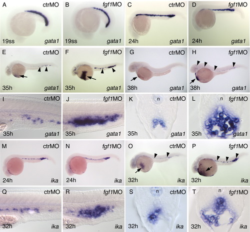
fgf1 morphants show altered gata1 and ika expression. Labeling of panels is as in Figure 2>. A-D: Early gata1 expression is normal in fgf1 morphants. E-H: Strong and persistent gata1-expressing cells in fgf1 morphants. Arrows point to the blood sinus covering the yolk, arrowheads to gata1-expressing cells in the intermediate cell mass (ICM) or elsewhere in the embryo. I-L: Close-ups and sections through the posterior ICM at 35 hours postfertilization (hpf; gata1). M,N:ika expression is normal in fgf1 morphants at 24 hpf. O,P: At 32 hpf, strong ika expression persists in fgf1 morphants, whereas it is down-regulated in control or wild-type embryos. Arrows/arrowheads as in E-H. Q-T: Cells strongly expressing ika accumulate in the ICM of fgf1 morphants. Close-ups and sections through the posterior ICM at 32 hpf. n, notochord
|