
Fig. 6
- ID
- ZDB-FIG-070309-22
- Publication
- Svetic et al., 2007 - Sdf1a patterns zebrafish melanophores and links the somite and melanophore pattern defects in choker mutants
- Other Figures
- All Figure Page
- Back to All Figure Page
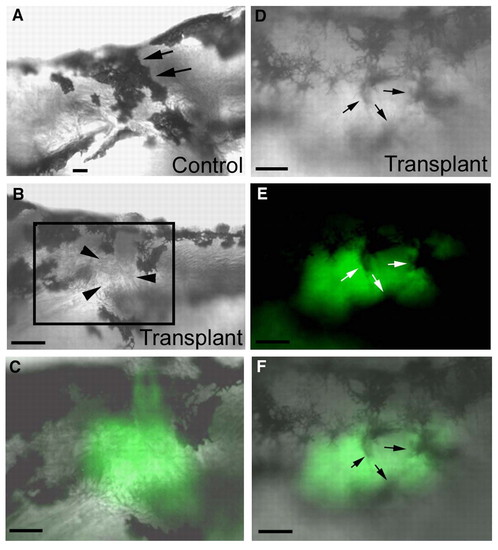
The cho mutant melanophore collar defects are caused by an underlying somite defect. (A-C) WT → cho mutant somite transplant embryo. (A) Right-hand, non-transplanted control side of a cho homozygote showing the melanophore collar at 5 dpf (arrows). Dorsal is to the top, anterior to the right. (B) Left-hand, experimental, transplanted side of the same embryo at the same time as in A. Note the gap in the collar on this side (arrowheads). (C) High magnification of boxed region in B, showing tight correlation of transplanted WT somite from alpha actin-GFP donor (green) with the collar region avoided by melanophores. Anterior is to the left, dorsal to the top. (D-F) The same transplanted WT → cho chimaeric embryo at 24 hours post-transplantation (2 dpf), showing that at early stages melanophores (arrows) traverse the transplanted tissue freely. Lateral views, anterior to the left, dorsal to the top. (D) Collar region on experimental side showing multiple melanophores traversing the transplant. (E) Fluorescent image showing the WT transplanted tissue (green). (F) D and E merged. Scale bars: 130 μm in B; 60 μm in A,C-F. |