|
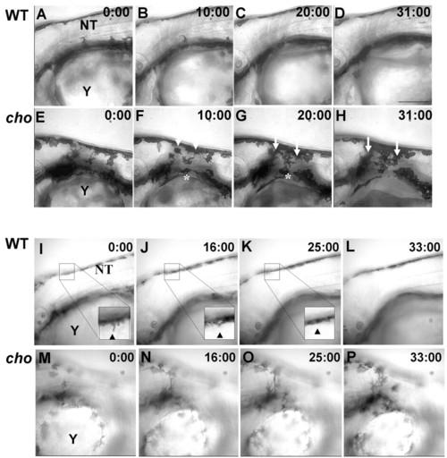
Timelapse microscopy of 48 hpf WT and cho mutants. (A-H) Images from different focal planes combined by projection revealed that WT melanophores (A-D) never entered the anterior trunk lateral pathway. By contrast, cho mutant melanophores (E-H) invaded the collar, both as single cells from the ventral stripe (asterisk) and as a sheet from the dorsal stripe (arrows). (I-P) Images from single focal planes clarify movements of individual cells. WT melanophores (I-L) extend processes into the anterior trunk (arrowhead, I) but these retract (arrow, arrowhead in J,K), whilst cho mutant melanophores (M-P) invade the collar, usually remaining ectopically. NT, neural tube; Y, yolk. Scale bar: 150 μm.
|