
Fig. 6
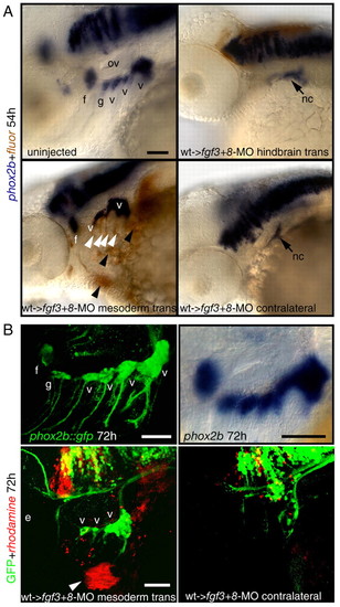
Restoration of cephalic mesoderm is sufficient to rescue EB ganglia. Wild-type donor embryos were injected with a lineage tracer fluorescein (A, brown), or rhodamine (B, red). At 30-40% epiboly, 25-30 donor cells were transplanted into the margin of fgf3+8-MO hosts. Mosaic embryos were collected at 54 (A) or 72 (B) hpf and analyzed for phox2b (blue, A) or GFP expression (green, B), respectively. (A) Lateral views of an uninjected control embryo (top, left) or fgf3+8 hosts that received hindbrain or mesodermal transplant. Note that mesodermal wild-type donor cells (brown stain, black arrowheads) efficiently rescued facial and vagal ganglia when compared with the contralateral control. Some donor cells also contributed to pharyngeal endoderm (white arrowheads). (B) phox2b::egfp transgene (top, left) recapitulates phox2b expression in the EB ganglia when compared with the endogenous message (top, right). Lateral view of the 72-hour old mosaic phox2b::egfp embryo that showed rescue of the vagal ganglia (compare with the contralateral side on the right). Donor cells (red, arrowhead) contributed to the facial muscles, indicating mesodermal origin. Note that in this example donor cells did not contribute to the endodermal pouches. Scale bars: 50 μm. Abbreviations are as in Fig. 1; ov, otic vesicle. |