
Fig. 1
- ID
- ZDB-FIG-070301-1
- Publication
- Mizoguchi et al., 2006 - Fgf signaling negatively regulates Nodal-dependent endoderm induction in zebrafish
- Other Figures
- All Figure Page
- Back to All Figure Page
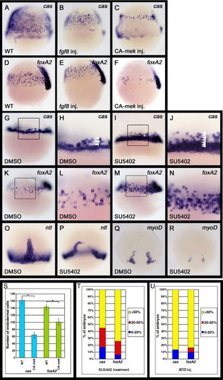
Fgf signaling can regulate the formation of endoderm and mesoderm. (A–F) Zebrafish embryos injected with fgf8 or CA-mek mRNA were examined for cas or foxA2 expression at the 60% epiboly stage (7 h postfertilization [hpf]). Lateral views, dorsal to the right. The number of cas- or foxA2-expressing cells is decreased in embryos in which Fgf signaling is activated. (G–N) Embryos treated with DMSO or SU5402 were examined for cas or foxA2 expression at either the 40% (5 hpf) or 60% epiboly stage (7 hpf), respectively. DMSO, SU5402: oep mRNA was injected into WT embryos at the one cell stage before treatment with DMSO or SU5402. (G–J) Lateral views, anterior to the top. (H and J) Enlarged views of the boxed area in panels G and I, respectively. Arrowheads in panels H and J indicate the number of cas-expressing cells from the margin. SU5402 treatment significantly increases the number of cas-expressing endodermal cells around the marginal domain. (K–N) Lateral views, dorsal to the right. (L and N) Enlarged views of the boxed areas in panels K and M, respectively. The number of foxA2-expressing endodermal cells is also significantly increased by SU5402 treatment. (O–R) Embryos treated with SU5402 were examined for expression of the pan-mesodermal marker ntl and adaxial maker myoD at the 80% epiboly stage (8. 3 hpf). The ntl and myoD expression levels were reduced by SU5402 treatment at the late stages of gastrulation. (S) Effects of the activation of Fgf signaling on the number of cas- or foxA2-expressing endodermal cells. The number of cas- or foxA2-expressing endodermal cells was counted at the 60% epiboly stage on both the left and right lateral sides of the embryos, which we determined to be representative of whole embryos. The embryos analyzed were cas-expressing endodermal cells in WT (n = 37) and CA-mek-injected (n = 45) embryos; foxA2-expressing endodermal cells in WT (n = 52); and CA-mek-injected (n = 38) embryos. Error bars represent the standard error. *P < 0.0005, Student′s t test. (T, U) Effects of the inhibition of Fgf signaling by SU5402 treatment (T) or XFD overexpression (U) upon the number of cas- or foxA2-expressing endodermal cells. oep mRNA was injected into WT embryos at the one cell stage before treatment with SU5402 (T) and was co-injected with XFD mRNA at the one cell stage (U). Zebrafish embryos at the 60% epiboly stage (7 hpf) were scored as demonstrating higher cas or foxA2 expression levels if they exhibited a greater than 50% (yellow), 20–50% (red) or 0–20% (blue) increase in endodermal cells. However, because SU5402 treatment or XFD overexpression greatly increases the number of cas- and foxA2-expressing endodermal cells, it is difficult to determine their exact number. The embryos analyzed were (T) cas-expressing endodermal cells in DMSO-treated (n = 37) and SU5402-treated (n = 48) embryos; foxA2-expressing endodermal cells in DMSO-treated (n = 52) and SU5402-treated (n = 51) embryos: (U) cas-expressing endodermal cells in WT (n = 28) and XFD-injected (n = 30) embryos; foxA2-expressing endodermal cells in WT (n = 33) and XFD-injected (n = 31) embryos. |
| Genes: | |
|---|---|
| Fish: | |
| Anatomical Term: | |
| Stage: | Shield |
Reprinted from Developmental Biology, 300(2), Mizoguchi, T., Izawa, T., Kuroiwa, A., and Kikuchi, Y., Fgf signaling negatively regulates Nodal-dependent endoderm induction in zebrafish, 612-622, Copyright (2006) with permission from Elsevier. Full text @ Dev. Biol.