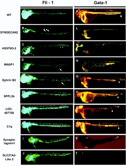
Defects in vasculogenesis and hematopoeisis observed in Tg (fli-1:eGFP (green)) or Tg (gata-1:DsRed (red)) embryos following MO inactivation of select CTT genes. (A) Normal vascular development observed in untreated Tg (fli-1:eGFP) embryos. (B, L) Decreases in the number of vascular sprouts (arrow heads) observed following injection of MO targeting Syndecan-2[20]. (C, M) Gaps within the caudal vein plexus (small arrow heads) observed following injection of MO targeting heparin sulfatetransferase-6-O 2-sulfotransferase (HSST6O-2)[21]. (D, N) Loss of integrity in the caudal vein plexus (arrow) observed following injection of MO targeting MAGP1[22]. Premature return in caudal vein flow shown by gata-1:dsRed expression to varying severities (arrowheads) following injection of MOs targeting Ephrin B2 (E,O), SPPL2b (F,P), predicted protein LOC407708 (G, Q), and C1q (H, R). Note: the premature return defects were not shown by fli-1:eGFP expression (E, F, G, H), however, were confirmed by other vascular markers (data not shown). (K) Normal blood development observed in untreated Tg (gata-1:DsRed) embryos. Decreased number of blood cells observed in 2 dpf embryos following injection with MO against Synaptotagmin13 (S) or Novel Protein similar to SLC27A2 (T). Accompanying panels (I) and (J) display no major vasculature defects for each of these genes respectively.
|
