
Fig. 5
- ID
- ZDB-FIG-070215-10
- Publication
- Phillips et al., 2006 - Zebrafish msxB, msxC and msxE function together to refine the neural-nonneural border and regulate cranial placodes and neural crest development
- Other Figures
- All Figure Page
- Back to All Figure Page
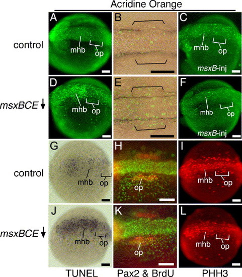
Cell division and cell death in msxBCE morphants. (A–F) Vital staining with acridine orange (AO) in wild-type embryos at 14 hpf (A) and 15 hpf (B), a 14 hpf wild-type embryo injected with msxB plasmid (C), msxBCE morphants at 14 hpf (D) and 15 hpf (E), and a 14 hpf msxBCE morphant injected with msxB plasmid (F). Images in panels B and E show transmitted light plus fluorescence to show otic placode morphology (brackets) in relation to AO-staining. (G, J) Wholemount TUNEL staining at 14 hpf in a control embryo (G) and msxBCE morphant (J). (H, K) Staining at 14 hpf for BrdU (green) and Pax2 (red) in a control embryo (H) and msxBCE morphant (K). (I, L) Staining at 14 hpf for Phosphohistone H3 (PHH3) in a control embryo (I) and msxBCE morphant (L). All images show dorsal or dorsolateral views with anterior to the left. Abbreviations: mhb, midbrain–hindbrain border; op, otic placode. Scale bars, 100 μm. |
| Gene: | |
|---|---|
| Fish: | |
| Knockdown Reagents: | |
| Anatomical Term: | |
| Stage: | 10-13 somites |
Reprinted from Developmental Biology, 294(2), Phillips, B.T., Kwon, H.J., Melton, C., Houghtaling, P., Fritz, A., and Riley, B.B., Zebrafish msxB, msxC and msxE function together to refine the neural-nonneural border and regulate cranial placodes and neural crest development, 376-390, Copyright (2006) with permission from Elsevier. Full text @ Dev. Biol.