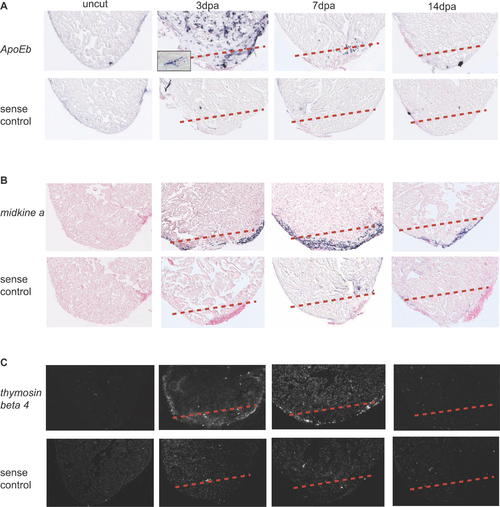
apoEb, midkine a, and thymosin β4 Are Expressed around the Wound Site during Zebrafish Heart Regeneration. Spatial expression patterns of apoEb, midkine a, and thymosin β4 in sham-operated hearts (uncut) and regenerating hearts at 3, 7, and 14 dpa were determined by in situ hybridization. (A) The expression pattern of apoEb was determined using a DIG-labeled antisense probe. apoEb was highly expressed at 3 dpa; its punctate expression suggests that it is expressed by infiltrating macrophages (see the higher magnification image in the inset). (B) Expression level of midkine a was upregulated from 3 dpa, reaches a peak at 7 dpa and lasts until 14 dpa. It appears to be expressed around the wound site in the compact layer of myocardium and the epicardium. (C) The expression pattern of thymosin β4 was determined using a radioactive antisense probe. thymosin β4 appears to be expressed around the wound and surrounding compact myocardium. Sense probes for each gene were used as negative controls. A dashed line marks the amputation plane.
|
