|
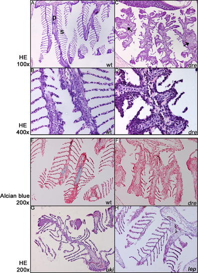
Patterning of the Branchial Arches in a 5-Mo-Old dre Mutant
(A and B) The strict organization of the brachial arch into primary (p) and secondary (s) lamellae in a wild-type situation (100× magnification). Higher magnification shows stacks of single chondrocytes in the primary lamellae.
(C and D) Sectioning of a dre mutant shows disturbed patterning, resulting in the absence of secondary lamellae and the presence of foci of chondrocyte-like cells in the primary lamellae (arrowsHE, hematoxylin and eosin stain; wt, wild-type.
(E and F) Alcian Blue staining reveals the presence of differentiated chondrocytes in the wild-type (wt) primary lamellae, but not in the dre mutant, indicating that the differentiation of these chondrocytes is affected (200×).
(G and H) Branchial arches of uki and lep mutants appear to be wild-type (wt).
|