|
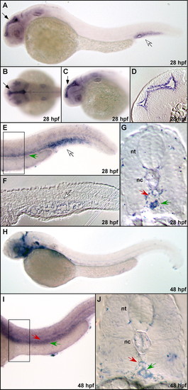
In-situ analysis of vtn mRNA expression. The vtn mRNA is robustly expressed in the ventricular zone (black arrow) at 28 hpf (A-C), also shown in section in D. Expression is also strong in the intermediate cell mass (A,E,F, white arrow). Lengthened staining also reveals vtn message in the vein (E, green arrow) confirmed by sectioning (G). At 48 hpf, vtn is expressed in the brain and diffusely in the branchial arches (H). Expression in the vein is detectable by whole-mount (I) and from sections (J), but is weaker than expression of san at this stage. The position of the dorsal aorta is indicated by the red arrow. The boxed region in E and I represent the region shown in the sections. nt, neural tube; nc, notochord.
|