|
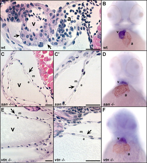
Morphological analysis of cardiac chambers of san and vtn mutant embryos. Hematoxylin- and eosin-stained sagittal sections through 72 hpf hearts were used to assess cardiac histology (A,C,E). Double-staining using an atrial-specific antibody (S46) and an in-situ RNA probe to the ventricular myosin heavy chain (vMHC) were used to distinguish the atrial and ventricular chambers (B,D,F). The myocardial wall (indicated by arrows) in wild-type hearts (A) is several cell layers thick. However, the myocardium in both san (C') and vtn (E') mutant hearts does not thicken and remains a single cell layer. Both mutants are shown at a reduced magnification in C and E to illustrate the dramatic dilation of the cardiac chambers. The hearts of (B) wild-type, (D) san-/-, and (F) vtn-/- embryos at 48 hpf express markers specific for the two cardiac chambers although the hearts of both mutants are enlarged. Scale bars: 25 μm. a, atrium; e, endocardium; m, myocardium; v, ventricle.
|