
Fig. 7
- ID
- ZDB-FIG-060808-243
- Publication
- Stigloher et al., 2006 - Segregation of telencephalic and eye-field identities inside the zebrafish forebrain territory is controlled by Rx3
- Other Figures
- All Figure Page
- Back to All Figure Page
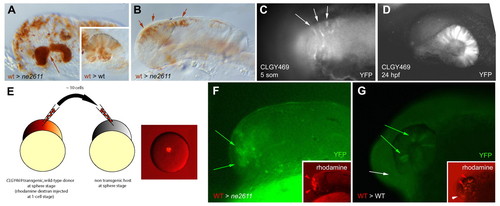
Rx3 controls eye field versus telencephalic fate in a cell-autonomous manner. (A,B) Transplantation of wild-type cells (brown) into the anterior forebrain of a ckhne2611 or wild-type (inset) host. (A) When a large number of cells are transplanted, retinal structures are rescued and evaginate (arrow). They are entirely composed of wild-type cells, whereas transplanted cells distribute randomly in a wild-type host (inset). (B) When a low number of cells is transplanted, retinal evagination does not occur. (C,D) Expression of YFP (revealed by anti-GFP immunocytochemistry) in transgenic CLGY469 embryos before (C; dorsal view) and after (D; lateral view) retinal evagination. YFP expression is restricted to eye-field cells and their descendants. It is absent in ckhne2611 mutants (see text). (E-G) Transplantation of a few wild-type cells transgenic for CLGY469 into the animal pole of a ckhne2611 (F) or wild-type (G) host. In a mutant host (F), retinal evagination does not occur (as in B), but expression of the transgene (green) is rescued in some transplanted cells (red, inset), indicating rescue of the eye-field fate. In a wild-type host, transplanted cells contribute to the retina (red arrowheads) and telencephalon (white arrowhead; G, inset), but only retinal cells express the transgene (G, green arrows, as opposed to white arrow). |