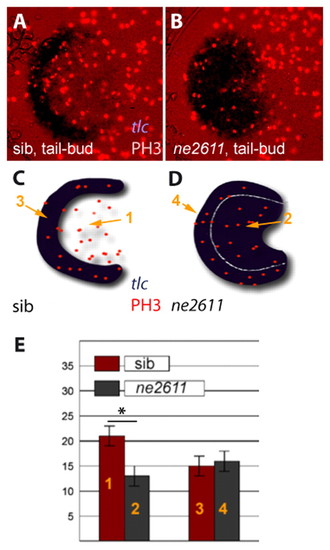
Rx3 accounts for the higher proliferation of eye field versus telencephalic precursors. (A,B) Immunodetection of the M-phase marker Phosphohistone-H3 (red nuclei) in the anterior neural plate of ckhne2611 mutants (right panel) and their wild-type siblings (left panel) at tail-bud stage (dorsal views, anterior left). tlc expression (ISH, blue) serves as a marker of the presumptive telencephalon in wild-type embryos and of the telencephalon+eye field in ckhne2611. (C,D) Schematic representation of the embryos in A,B to localize the presumptive telencephalic and eye fields in wild-type embryos (domains 3 and 1, respectively) and the corresponding territories in ckhne2611 siblings (domains 4 and 2, respectively) at tail-bud stage. (E) Number of PH3-positive cells in domains 1-4 (see C,D) in wild-type (red) versus ckhne2611 (gray) embryos. Bars indicate standard errors. Proliferation is significantly decreased in the presumptive eye field, but is unaltered in the presumptive telencephalon, in ckhne2611 compared with wild-type embryos at tail-bud stage (two-sample independent Student's t-test, P values are given in the text).
|
