
Fig. 3
- ID
- ZDB-FIG-060629-3
- Publication
- Wendik et al., 2004 - Zebrafish mnx genes in endocrine and exocrine pancreas formation
- Other Figures
- All Figure Page
- Back to All Figure Page
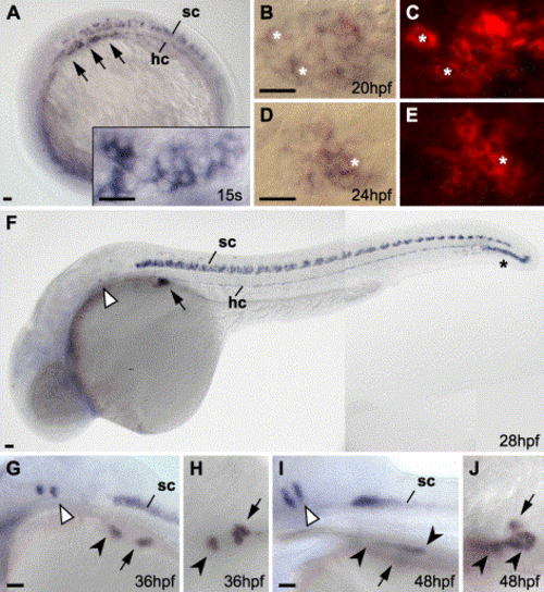
Tissue-specific expression of hb9 in endocrine pancreas. Expression of hb9 in purple at 15 somite (A), 20 hpf (B, C), 24 hpf (D, E), 28 hpf (F), 36 hpf (G, H) and 48 hpf (I, J). Between 15 somite stage (inlay in A shows a dorsal view of the same embryo in a higher magnification) and 24 hpf endodermal hb9 expression changes from an elongated mono-layered domain to a condensed structure underlying the hypochord (hc). (B–E) Bright field (B, D) and fluorescence (C, E) images of double in situ stains for hb9 (NBT/BCIP) and insulin (Fast Red) show expression of insulin in most hb9-expressing cells; asterisks mark examples for double-labeled cells. Further indicated are hb9 expression domains in the spinal cord (sc), rhombomeres 5 and 6 (white arrowheads) and the swim bladder (black arrowheads). Embryos are shown from a lateral (A, F, G, I) or dorsal view (B, C, D, E, H, J) with anterior to the left. Scale bars correspond to 20 μm. |
| Genes: | |
|---|---|
| Fish: | |
| Anatomical Terms: | |
| Stage Range: | 14-19 somites to Long-pec |
Reprinted from Developmental Biology, 268(2), Wendik, B., Maier, E., and Meyer, D., Zebrafish mnx genes in endocrine and exocrine pancreas formation, 372-383, Copyright (2004) with permission from Elsevier. Full text @ Dev. Biol.