
Fig. 6
- ID
- ZDB-FIG-060424-10
- Publication
- Grinblat et al., 1998 - Determination of the zebrafish forebrain: induction and patterning
- Other Figures
- All Figure Page
- Back to All Figure Page
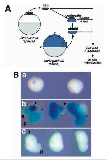
Early gastrula organizer (shield) induces oplexpression in animal caps. (A) Schematic outline of the conjugation experiment. Animal cap explants from late blastula (sphere stage) embryos were cultured either in isolation, in groups of 10, or as conjugates with shields explanted from early gastrula (shield stage) embryos. Each conjugate was made with one shield and 5 animal caps. As a control, shields were cultured alone. For conjugates, embryos from which shields were isolated were lineage labeled with FLDX (pale blue; see Methods) in order to distinguish inducing from responding tissues. Explants were cultured until shield-donor embryos reached the 5 somite stage, at which time the explants were harvested and stained for opl RNA (purple) by in situ hybridization. (B) Cultured explants, stained for the presence of opl RNA (purple) and lineage label (blue). (a) Two groups of 10 animal caps cultured in isolation. (b) Three conjugates representative of the typical outcomes of the experiment. Dots mark the anterior and posterior edges of the animal cap-derived tissues. (c) Shield explants cultured in isolation. A total of 38 conjugates were made in 7 independent experiments. Induction of oplwas observed in 58% of them, and was always restricted to the animal cap-derived portion. No detectable oplexpression has been observed in more than 40 shield explants generated in at least 5 experiments. Abbreviations: a, anterior; p, posterior. |