
Fig. 5
- ID
- ZDB-FIG-060307-6
- Publication
- Sakai et al., 2006 - Semaphorin 3d guides laterality of retinal ganglion cell projections in zebrafish
- Other Figures
- All Figure Page
- Back to All Figure Page
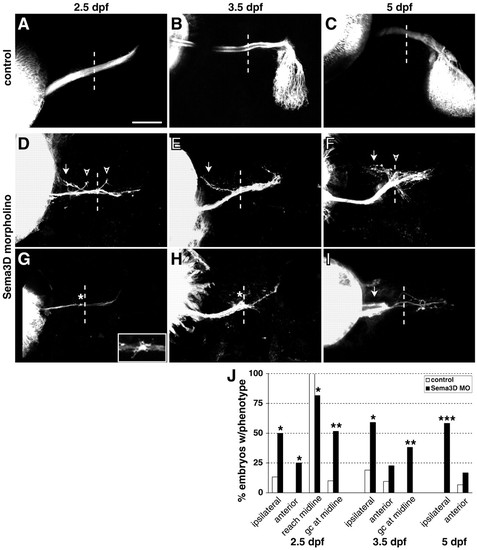
Sema3d knockdown increases ipsilateral guidance errors and reduces midline crossing. (A-I) Confocal projections of DiI-labeled RGC axons in control (A-C) or Sema3d (D-I) morpholino-injected embryos at 2.5 (A,D,G), 3.5 (B,E,H) and 5 (C,F,I) dpf; injected eyes are on the left. A,D-I are ventral views; B and C are dorsal views. Sema3d knockdown caused aberrant projections ipsilaterally (arrows) and anteriorly (arrowheads) from the normal pathway, and increased the occurrence of axons extending to but not beyond the midline (asterisks). Inset in G shows an enlargement of a growth cone near midline. Dotted lines indicate midlines; anterior is up in all panels. Scale bars: 50 μm in A,D-I; 80 μm in B,C. (J) Summary of guidance and extension errors following Sema3d morpholino (Sema3d MO) knockdown. At 2.5 dpf: control morpholino, n=30 for all analyses; Sema3d morpholino, n=38 analyzed for reaching the midline, n=31 analyzed for presence of growth cones, n=28 analyzed for guidance errors. At 3.5 dpf: control morpholino, n=21; Sema3d morpholino, n=22. At 5 dpf: control morpholino, n=30; Sema3d morpholino, n=36. *P<0.01; **P<0.001; ***P<0.000001. |