
Fig. 2
- ID
- ZDB-FIG-060307-3
- Publication
- Sakai et al., 2006 - Semaphorin 3d guides laterality of retinal ganglion cell projections in zebrafish
- Other Figures
- All Figure Page
- Back to All Figure Page
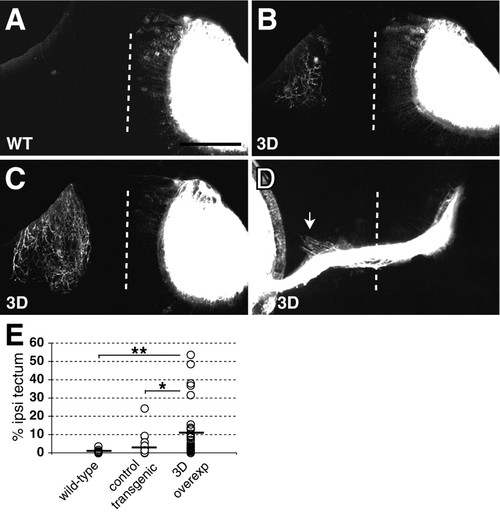
Ubiquitous Sema3d overexpression increases aberrant ipsilateral projections. (A-C) Confocal projections of dorsal views of tecta in 5 dpf wild-type (A) and Sema3d-overexpressing (B,C) embryos show tectal innervation. Left eyes were injected and labeled eyes were digitally removed from images. Wild-type embryos (WT) contained few or no ipsilateral axons (A), whereas Sema3d-overexpressing embryos (3D) contained more extensive ipsilateral arbors (B,C). (D) Confocal projection of a ventral view of the optic chiasm in a 3 dpf Sema3d-overexpressing embryo, injected eye on the left, shows ipsilaterally projecting axons in the region of the optic tract (arrow). Ipsilateral axons are obscured near the midline by the labeled optic nerve in this projection. Dotted lines indicate midlines; anterior is up in all panels. Scale bars: 100 μm in A-C; 63 μm in D. (E) Scatter plot summarizing the extent of ipsilateral tectal innervation in each embryo at 5 dpf; lines indicate means. Sema3d overexpression significantly increased ipsilateral innervation at 5 dpf. WT, n=25; control transgenic embryos, n=20; Sema3d-overexpressing (3D overexp) embryos, n=32. *P<0.05, **P<0.02. |