
Fig. S2
- ID
- ZDB-FIG-060307-13
- Publication
- Rentzsch et al., 2006 - Crossveinless 2 is an essential positive feedback regulator of Bmp signaling during zebrafish gastrulation
- Other Figures
- All Figure Page
- Back to All Figure Page
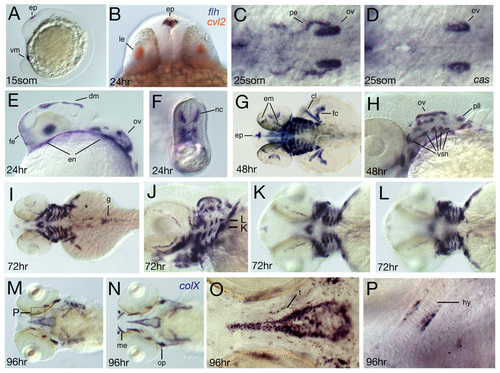
Spatial expression of crossveinless2 at later developmental stages. Developmental stage is indicated in the lower left corner. (A, C-M, O-S) In situ hybridization with a cvl2 antisense probe, (B) double in situ staining with cvl2 in red and floating head (flh) in blue, and (N) in situ hybridization with a collagen X probe. (A,E) Lateral views, (B) frontal optical cross-section showing co-expression of cvl2 and flh in the epiphysis. (C,D) Dorsal view of (C) wild type and (D) casanova mutant. Expression in the pharyngeal endoderm is absent in casanova mutants, (F) optical cross-section of the tail at the level of the yolk extension showing expression in medially migrating neural crest cells, (G,I,K,L) dorsal views, focal planes of K,L are indicated in J. (K) Expression in putative medioventral cartilage condensations (see also J). (L) Expression in more dorsolateral areas that separate the arches (compare also with J). (M-P) Ventral views, expression domains of cvl2 and the bmp target gene collagen X overlap. (O) Higher magnification of the area of the hypophyseal fenestre showing that cvl2 is not expressed in the stacked mature chondrocytes of the trabeculae. (P) Higher magnification of the hyoid arch as indicated in M. cvl2 expression is restricted to tissue surrounding the cartilage. cl, cleithrum; dm, dorsal diencephalic midline; em, eye muscles; en, endoderm; enp, endodermal pouches; ep, epiphysis; fc, finbud cartilage; fe, facial ectoderm; g, gut; hy, hyoid arch; le, lens; nc, neural crest; pe, pharyngeal endoderm; pll, posterior lateral line ganglion; op, operculum; ov, otic vesicle; t, trabeculae; vm, ventral mesoderm; vsn, visceral sensory neurons. |