
Fig. 3
- ID
- ZDB-FIG-060118-4
- Publication
- Pollard et al., 2006 - Essential and overlapping roles for laminin alpha chains in notochord and blood vessel formation
- Other Figures
- All Figure Page
- Back to All Figure Page
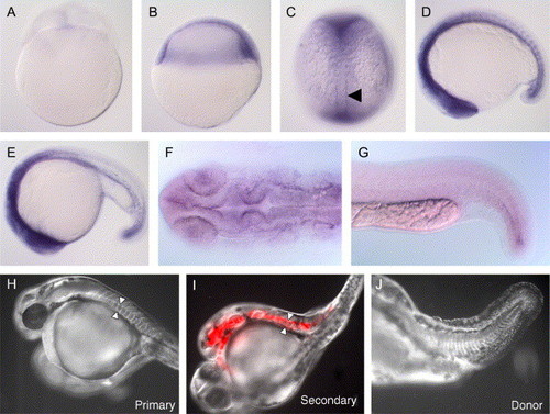
Expression of lama1 mRNA and genetic mosaic experiments reveal that laminin α1 is supplied from non-notochordal tissues. Whole-mount in situ hybridisation for lama1 mRNA in wild-type zebrafish embryos at: 8-cell stage (A), shield stage (B), 5-somite stage (C, dorsal view), 15-somite stage (D), 25-somite stage (E) and 24 hpf (F, dorsal view of head; G, lateral view of trunk and tail). Embryonic shield transplantation from a Rhodamine-dextran labelled bal mutant donor (J), into wild-type host (H), gives rise to a secondary axis containing a rescued anterior notochord (red fluorescence) (I). Morphologically wild-type notochord in both the primary (host) axis and secondary (donor) axis are indicated with white arrowheads. |
| Gene: | |
|---|---|
| Fish: | |
| Anatomical Terms: | |
| Stage Range: | 8-cell to Prim-5 |
Reprinted from Developmental Biology, 289(1), Pollard, S.M., Parsons, M.J., Kamei, M., Kettleborough, R.N., Thomas, K.A., Pham, V.N., Bae, M.K., Scott, A., Weinstein, B.M., and Stemple, D.L., Essential and overlapping roles for laminin alpha chains in notochord and blood vessel formation, 64-76, Copyright (2006) with permission from Elsevier. Full text @ Dev. Biol.