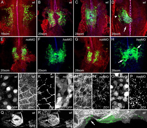
Myocardial epithelial organization is disrupted in has/prkci and nok/mpp5 morphants. All images represent reconstructions of confocal Z-stack sections imaged on whole mounts. (A-E,G) cmlc2:GFP, green (nuclear GFP within myocardial cells); PRKC/aPKC, red; ZO-1, blue. PRKC and ZO-1 were used as a counterstain to visualize the embryonic midline (see also dotted line in D). (F,H) cmlc2:GFP, green and ZO-1, blue. (Q-S) cmlc2:GFP and filamentous actin. (A) At the 16-somite stage, wild-type myocardial cells are organized into two bilateral sheets of cells. (B) Both sheets converge onto the midline, where they fuse to form the heart cone around the 20-somite stage. (C) Further convergence leads to a narrowing of the heart cone and expansion along the dorsoventral axis around the 24-somite stage. The entire heart cone moves out of the midline toward the left and anterior. (D) Heart cone tilting places the heart into the anterior-posterior orientation by the 28-somite stage. The atrium (arrowhead) is located to the left and anterior whereas the ventricle (arrow) is oriented toward the midline and posterior. (E,G) nok/mpp5 morphants display a delay in myocardial fusion to form the heart cone. (F,H) has/prkci morphants undergo heart cone fusion at the midline but tilting of the heart cone into the anterior-posterior orientation fails. Note the disrupted appearance of the myocardial layer, with holes and rough edges (arrows). (I-P) All images are details of confocal reconstructions of confocal Z-stacks imaged from 16-somite stage whole mounts, separated into the individual channels. (I-K) At the 16-somite stage, wild-type myocardial cells have an epithelial organization with junctional distribution of PRKC and ZO-1. (L-N) At the 16-somite stage, nok/mpp5 morphants display a diffuse distribution of PRKC along membranes and ZO-1 junctional belts are disrupted and appear as spots. (O,P) At the 16-somite stage, has/prkci morphants display a spotted distribution of ZO-1. (Q,R) Dorsal view onto the heart cone shown in S. (S) During tilting, specialized elongated myocardial cells containing actin-cables attach the heart cone to surrounding tissues (see arrow). Orientation: dorsal view and anterior to the top, except in S, lateral view and anterior to the left.
|
