|
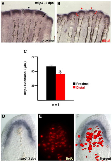
Position-dependent length of Fgf target gene expression domains. (A,B) Images from the same double-amputated fin regenerate demonstrates a longer PD length of mkp3 expression (asterisks, brackets) in proximal regenerates. (C) The length of the proximal signal was 28% longer than the distal signal on average (n=8; *P<0.005, t-test). (D-F) 3 dpa fin regenerate (33°C) stained for mkp3 expression (D) and BrdU incorporation (E). Cells in the distal blastema and basal epidermal layers expressing mkp3 show little proliferation. However, proliferative blastemal mesenchyme is bordered by epidermal mkp3 expression/Fgf signaling (F).
|