
Fig. 1
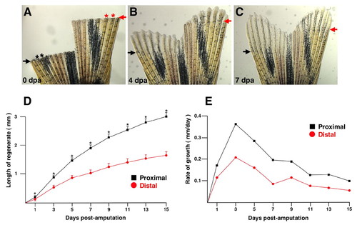
Amputated zebrafish caudal fins display position-dependent rates of regenerative growth. (A) Appearance of the zebrafish fin immediately following a double amputation surgery, with the injured portion at the top of the image. The amputation planes are indicated by arrows (black, proximal; red, distal), and asterisks mark lateral rays 2 and 3 that are compared in this study. (B) Only 4 days after amputation (dpa; assessed at 33°C), the fin has regenerated a significant number of lost structures. The ventral lobe of the fin (left), after a more proximal amputation, is regenerating more rapidly than the right, dorsal lobe. (C) By 7 dpa, the ventral regenerate has reached nearly the same PD level as the dorsal regenerate. (D) Growth is greater after proximal amputations compared with distal amputations throughout regeneration (mean±s.e.m.; *P<0.05, t-test). (E) Growth rate is greater after proximal amputations than distal throughout regeneration. |