
Fig. 2
- ID
- ZDB-FIG-051220-2
- Publication
- Ando et al., 2005 - Lhx2 mediates the activity of Six3 in zebrafish forebrain growth
- Other Figures
- All Figure Page
- Back to All Figure Page
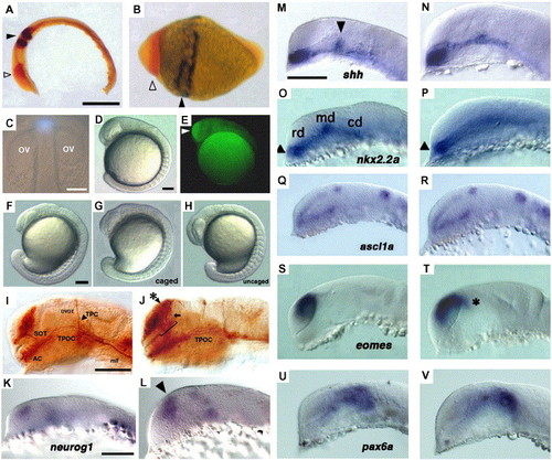
Comparison of the effect of ubiquitous Lhx2 overexpression with that of head-specific photoactivation of caged-lhx2 mRNA. (A) Two-color in situ hybridization of an uninjected embryo at 15 hpf. Red signal shows pax6a expression in the head, and dark stripes show egr2b expression in the hindbrain. (B) Two-color in situ hybridization in the Lhx2-overexpressing embryo at 15 hpf. Embryo showed severe dorsalization as well as anteriorization at this stage. Open arrowhead points to the expanded region of pax6a expression in the enlarged head, and closed arrowhead points to the expanded region of egr2b expression. (A, B) Lateral view, anterior left and dorsal up. Scale bar: 200 μm. (C–H) Uncaging by spot UV illumination induced head-specific overexpression of Lhx2:GFP fusion protein. (C) Spot UV illumination, 36 μm in diameter, with 365-nm light (shown as blue spot) of the presumptive forebrain of a 12-hpf embryo to uncage lhx2:gfp fusion mRNA. Illumination was provided by a 100-W mercury lamp for 1.0 s using a 10-fold objective lens and a 200-μm pinhole slide inserted at the confocal plane of the light pathway. ov, optic vesicles. (D) Head-uncaged embryo at 16 hpf. (E) Fluorescence microscopy image of the head-uncaged embryo shown in panel D. GFP fluorescence was only detected in the head (white arrowhead). (F–H) Morphology of embryos at 18 hpf. (F) An uninjected embryo. (G) An embryo injected with caged-fusion mRNA without uncaging treatment. (H) An embryo injected with caged-fusion mRNA and uncaged in the head at 12 hpf. (I, J) Immunostaining of the primary neurons with α-Tb of a 24-hpf uninjected (I) and head-uncaged embryo (J). Asterisk in panel J points to the ectopic neurons in the dorsal telencephalon that extend their axons in the defasciculated supraoptic tracts (arrow and bracket) toward the rostral TPOC. TPOC, tract of the postoptic commissure; SOT, supraoptic tract; TPC, tract of the posterior commissure; DVDT, dorsoventral diencephalic tract; AC, anterior commissure; mlf, medial longitudinal fascicle. (K, L) neurog1 expression at 17 hpf in uninjected (K) and head-uncaged embryo (L). Arrowhead in panel L shows expanded neurog1 expression. Scale bar, 100 μm. (M–V) Comparison of the expression patterns of brain marker genes in the uninjected (M, O, Q, S, U) and in the head-specific lhx2 mRNA-uncaged embryos (N, P, R, T, V). (M, N) shh expression at the 18-somite stage. Arrowhead in panel M points to dorsal shift of shh expression domain at the mid–diencephalic boundary. (O, P) nkx2.2a expression at the 18-somite stage. Arrowheads point to the optic recess of each embryo. rd, md, cd, rostral domain, medial domain, caudal domain of nkx2.2a expression in the uninjected embryo. (Q, R) ascl1a expression at 20 hpf. (S, T) eomes expression at 18 hpf. Asterisk in panel T points to dorsal extension of eomes expression. (U, V) pax6a expression at 24 hpf. Scale bar (M–V), 100 μm. |
| Genes: | |
|---|---|
| Fish: | |
| Anatomical Terms: | |
| Stage Range: | 10-13 somites to Prim-5 |
Reprinted from Developmental Biology, 287(2), Ando, H., Kobayashi, M., Tsubokawa, T., Uyemura, K., Furuta, T., and Okamoto, H., Lhx2 mediates the activity of Six3 in zebrafish forebrain growth, 456-468, Copyright (2005) with permission from Elsevier. Full text @ Dev. Biol.