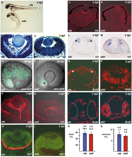
Retinal mitotic cells fail to exit from the cell cycle in add mutant embryos. (A) Morphology of add mutant and wild-type sibling embryos. (B,C) Plastic sections of wild-type (B) and add mutant (C) retinas. add mutant embryos show a markedly multifolded retinal epithelium. (D,E) ath5:GFP expression in wild-type (D) and add mutant (E) retinas. ath5:GFP expression is markedly delayed in add mutant embryos and very faint in the ventronasal retina (E, asterisk). (F,G) Labeling of wild-type (F) and add mutant (G) retinas with the zn5 antibody, which stains retinal ganglion cells (red). (H,I) Labeling of wild-type (H) and add mutant (I) retinas with the zpr1 antibody, which stains double cone photoreceptors (red). All nuclei were counterlabeled with Sytox Green (green). (J,K) Labeling of wild-type (J) and add mutant (K) retinas with the anti-glutamine synthetase antibody, which stains Müller glial cells (red). (L,M) In situ hybridization of wild-type (L) and add mutant (M) retinas with rx1 RNA probe. rx1 expression is observed in nearly the entire neural retina of add mutants, whereas it is downregulated and localized within a proliferating region called the CMZ (L, asterisks) in wild type. (N,O) BrdU labeling of wild-type (N) and add mutant (O) retinas. BrdU-positive cells are located within the CMZ (N, asterisks) and a part of the outer photoreceptor layer in wild type, whereas many retinal cells incorporate BrdU even in the central retina of add mutant embryo. (P,Q) BrdU labeling of wild-type (P) and add mutant (Q) heads. Broken white lines show the interface between the brain and retina at 24 hpf. (R) The percentage of the number of BrdU-positive cells to total number of cells in wild-type (blue bar) and add mutant (red bar) retinas. (S) The percentage of the number of mitotic cells labeled with anti-phosphorylated Histone H3 antibody in wild-type (blue bar) and add mutant (red bar) retinas at 27 hpf.
|
