FIGURE

Fig. 4

ID
ZDB-FIG-051102-4
Publication
Kaji et al., 2004 - dlx3b and dlx4b function in the development of Rohon-Beard sensory neurons and trigeminal placode in the zebrafish neurula
Other Figures
All Figure Page
Back to All Figure Page
Fig. 4

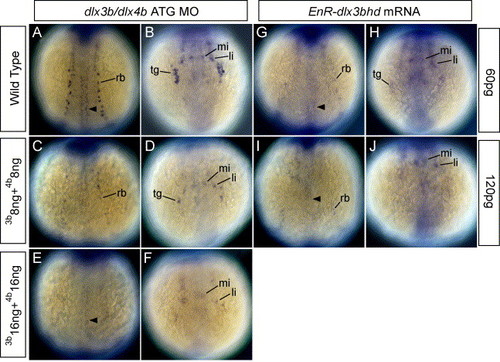
HuC expression in embryos injected with dlx3b-ATG-MO and dlx4b-ATG-MO and EnR-dlx3bhd mRNA at 3-somite stage. (A, C, E, G, I) Dorsal views, anterior is to the top. (B, D, F, H, J) Anterior views. (A and B) Wild-type embryos. (C and D) 8 ng dlx3b-MO- and 8 ng dlx4b-MO-injected embryos. (E and F) 16 ng dlx3b-MO- and 16 ng dlx4b-MO-injected embryos. (G and H) 60 pg EnR-dlx3bhd mRNA-injected embryos. (I and J) 120 pg EnR-dlx3bhd mRNA-injected embryos. (A–F) and (G–J) show that the expression of HuC, which is expressed in RB neurons in the trunk (rb) and neurons in trigeminal placodes in the head (tg), is gradually reduced as the concentration of MOs and EnR-dlx3bhd mRNA are increased. In contrast, primary motor neurons (arrowhead) and anterior lateral hindbrain interneurons (li) and anterior medial hindbrain interneurons (mi) are not changed.

Expression Data
Gene:
Fish:
Knockdown Reagents:
Anatomical Terms:
Stage: 1-4 somites

Expression Detail
Antibody Labeling
Phenotype Data
Fish:
Knockdown Reagents:
Observed In:
Stage: 1-4 somites

Phenotype Detail
Acknowledgments
This image is the copyrighted work of the attributed author or publisher, and ZFIN has permission only to display this image to its users. Additional permissions should be obtained from the applicable author or publisher of the image.

Reprinted from Developmental Biology, 276(2), Kaji, T., and Artinger, K.B., dlx3b and dlx4b function in the development of Rohon-Beard sensory neurons and trigeminal placode in the zebrafish neurula, 523-540, Copyright (2004) with permission from Elsevier. Full text @ Dev. Biol.