
Fig. 8
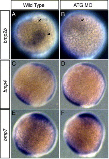
Expression of bmp2b, 4, and 7 in embryos injected with dlx3b-MO and dlx4b-MO at 90% epiboly. Expression in wild-type embryos of bmp2b (A), bmp4 (C), and bmp7 (E). Expression in 20 ng ATG-dlx3b-MO/20 ng ATG-dlx4b-MO-injected embryos of bmp2b (B), bmp4 (D), and bmp7 (F). (A and B) show that there is a strong triangle of bmp2b expression (arrowhead) at the border of the non-neural ectoderm in the wild-type embryos but this region is lost in MOs-injected embryos. Ventral bmp2b expression persisted in MOs-injected embryos (arrow). No alterations are seen in the expression of bmp4 and bmp7 between wild-type embryos and MOs-injected embryos (C–F). Lateral views, and dorsal is to the right. |
| Genes: | |
|---|---|
| Fish: | |
| Knockdown Reagents: | |
| Anatomical Term: | |
| Stage: | 90%-epiboly |
Reprinted from Developmental Biology, 276(2), Kaji, T., and Artinger, K.B., dlx3b and dlx4b function in the development of Rohon-Beard sensory neurons and trigeminal placode in the zebrafish neurula, 523-540, Copyright (2004) with permission from Elsevier. Full text @ Dev. Biol.