
Fig. 6
- ID
- ZDB-FIG-051025-6
- Publication
- Flanagan-Steet et al., 2005 - Neuromuscular synapses can form in vivo by incorporation of initially aneural postsynaptic specializations
- Other Figures
- All Figure Page
- Back to All Figure Page
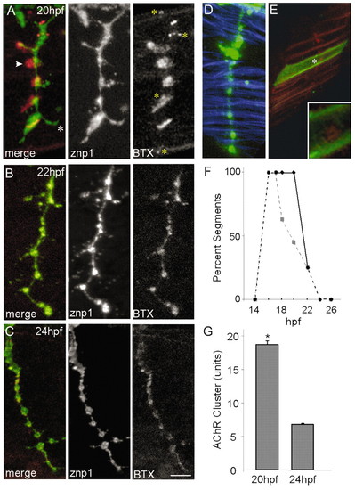
Apposition of CaP axon varicosities to AChR clusters. (A-C) Axons and AChRs labeled with znp-1 (green) and BTX (red), respectively. By 20 hpf, axonal varicosities are apposed to AChR clusters, but some clusters are larger than varicosities (arrowhead) and some axonal processes extend beyond clusters (asterisk). Yellow asterisks indicate AChR clusters that are not innervated. Apposition becomes precise by 24 hpf (B,C). At this time, pre- and postsynaptic specializations are perfectly aligned (C), varicosities are centered on myotubes (D; phalloidin in blue) and each myotube bears a single AChR cluster (E; single myotube labeled with mYFP by mosaic expression of a transgene injected into a one-cell embryo). The inset in E is a higher-power view of the single cluster indicated by the asterisk. (F) Progressive loss of aneural clusters. Aneural clusters lying rostral or caudal to CaP disperse (solid line), whereas many of the aneural clusters in front of the axon (dashed line) are incorporated into NMJs (n=10-64, average=38, segments per stage). (G) Cluster size decreases between 20 and 24 hpf (P<0.0001 by t-test; n=250 clusters per stage). Scale bar in C: 5 µm for A-E. |