
Fig. 2
- ID
- ZDB-FIG-051025-2
- Publication
- Flanagan-Steet et al., 2005 - Neuromuscular synapses can form in vivo by incorporation of initially aneural postsynaptic specializations
- Other Figures
- All Figure Page
- Back to All Figure Page
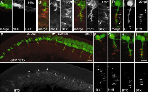
AChR clustering precedes axon outgrowth. CaP axons (green) labeled with znp-1 (B,C) or GFP (A,D-I) as in Fig. 1; AChRs labeled with BTX (red). (A-D) Segments 1-3. Neither axons nor AChR clusters are present at 14 hpf (A), but clusters are present in front of the advancing axon by 16-17 hpf (B,C). By 20 hpf, (D) most AChR clusters are apposed by axonal varicosities, but some clusters are larger than varicosities (arrowheads) and some axonal processes extend beyond clusters (arrow). Asterisks in B-D indicate some of the AChR clusters that are not innervated; they are quantified in Fig. 6F. (E-I) Whole mount of a 20 hpf fish (E), and higher-power details of individual segments (F-I) showing the rostrocaudal developmental sequence (see also Fig. 1A). In caudal segments (F), AChR clusters have formed but no axon has yet extended. In intermediate segments (G,H), clusters on adaxial cells not contacted by CaP disperse (asterisks in E). In the most rostral segments (I), the CaP axon has extended beyond the ventral-most AChR clusters. Scale bars: in A, 10 µm for A-D; in E, 25 µm; in I, 10 µm for F-I. |