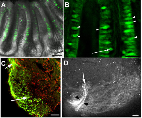
Expression of Rag1 in the adult zebrafish olfactory system.(A) An olfactory rosette isolated from a 1.5 year-old adult. GFP-expressing cells are located in the sensory region of the lamella and midline raphe. Most cell bodies are located apically. (B) High magnification of lamella from another olfactory rosette, also from a 1.5 year-old adult. GFP-expressing cells have differing morphologies. Many have the cell body close to the apical surface (arrowheads). The arrow indicates one neuron with a deep cell body. (C) Anti-GFP immunofluorescence on a horizontal section through the olfactory bulb of an adult. Label is detectable only in olfactory sensory neurons innervating the lateral bulb. The incoming olfactory nerve is visible at the upper left (arrow). Anterior is to the top-left; lateral to the bottom-left. Nuclei are labelled with propidium iodide (red). (D) Lateral view of an olfactory bulb dissected from a 1.5-year-old Rag1:GFP male. Axons innervate a large portion of the lateral bulb, but axons with the highest levels of GFP (black arrowheads) form two bundles that innervate a single region (arrow). Other glomeruli are innervated by dimmer axons. Bar = 50 µm (A, C, D); 20 µm (B).
|
